 by
by -
The OpenVertebrata Project, aka oVert, has CT scanned over 13,000 museum specimens.
-
Their newly published images allow you to see inside lizards, birds, rodents and more.
-
Over the next four years, the oVert team plans to CT scan another 20,000 museum specimens.
Have you ever wished you had X-ray vision?
The OpenVertebrate project, or oVert for short, is based on the next best thing: CT scanning.
Over the past six years, this project has partnered with 18 institutions to in-house scan over 13,000 museum specimens.
But they won’t stop there. The project team plans to ramp things up to 20,000 scans over the next four years.
Their 3-D images show the complex biodiversity of thousands of animal species.
“When people first collected these specimens, they had no idea what would become of them,” said Edward Stanley, the project’s co-principal investigator, in a press release.
The project’s mission is to improve the accessibility of museum specimens, and oVert has made their images publicly available so that researchers, and anyone curious about animal anatomy, can learn from them.
“If you want someone to get on a plane and travel to you to collaborate, that’s prohibitive in many ways,” said David Blackburn, the project’s principal investigator, in the press release.
“Now we have scientists, teachers, students and artists around the world using this data remotely,” Blackburn said.
A fruit bat with a black belly and huge wings
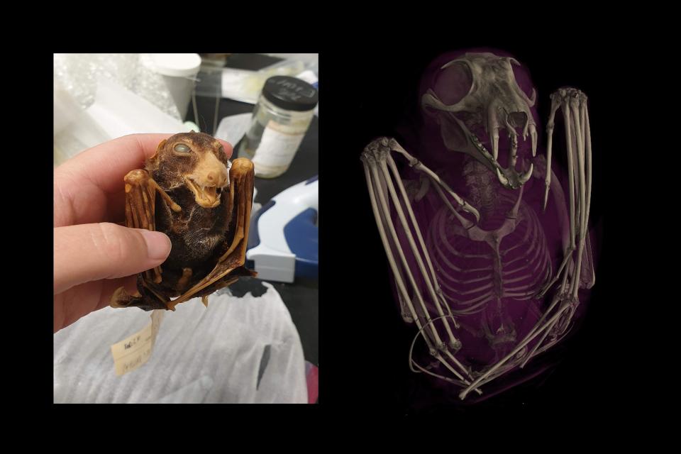
The images allow you to see animals like you’ve never seen them before, revealing their inner anatomy. Check out the skeletal structure of the wings of this black fruit bat.
A spiny mouse with an armored tail
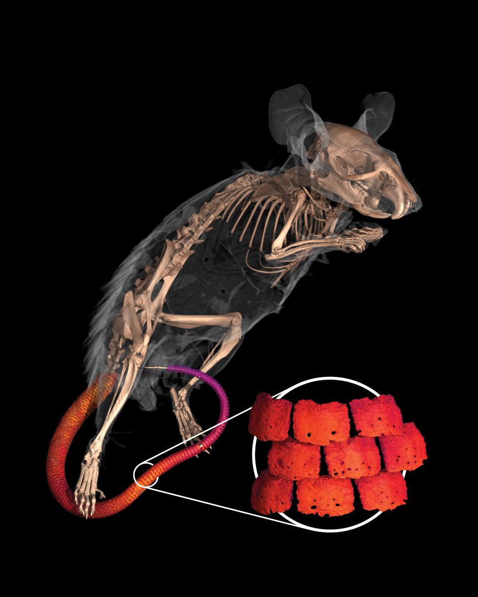
The project even made some new discoveries. A scan of this spiny mouse revealed its armored tail, helping to confirm that it is related to other rodents with similar characteristics.
A Mexican bearded lizard
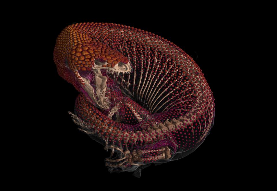
This Mexican bearded lizard is also suitable for armor. The oVert project allows scientists to trace the evolution of similar armor plating across fish, amphibians, reptiles, and even mammals.
A chameleon

This scan revealed the unique head shape of a veiled chameleon. The huge flask on top of his head is called a casque. Its main function is to put water droplets into the chameleon’s mouth.
A northern sheep frog with a big heart
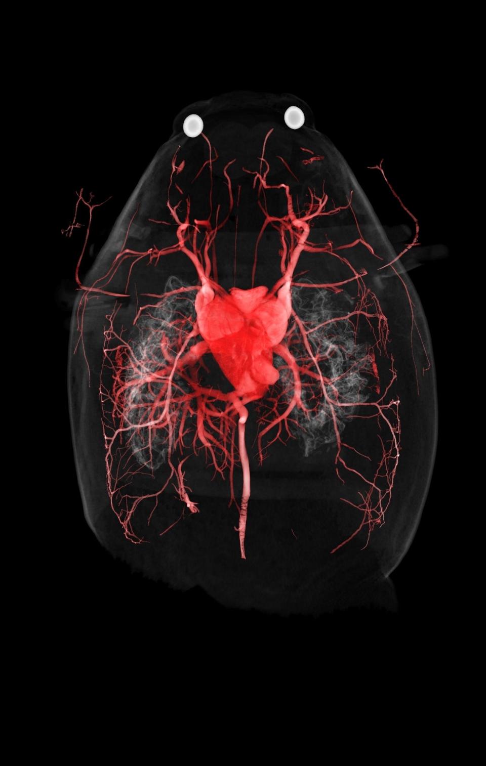
But the scans showed more than just bone structures. This is the vascular system of the Northern sheep frog. The heart is a large part of its body size.
Challenging echidna
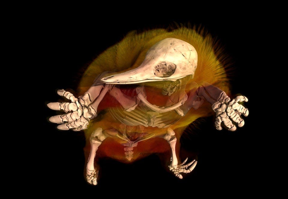
The most challenging specimen to scan was this echidna, due to its sharp spines. To create this image, oVert scientists had to secure the specimen in durable bags.
Complex skeleton of a seahorse
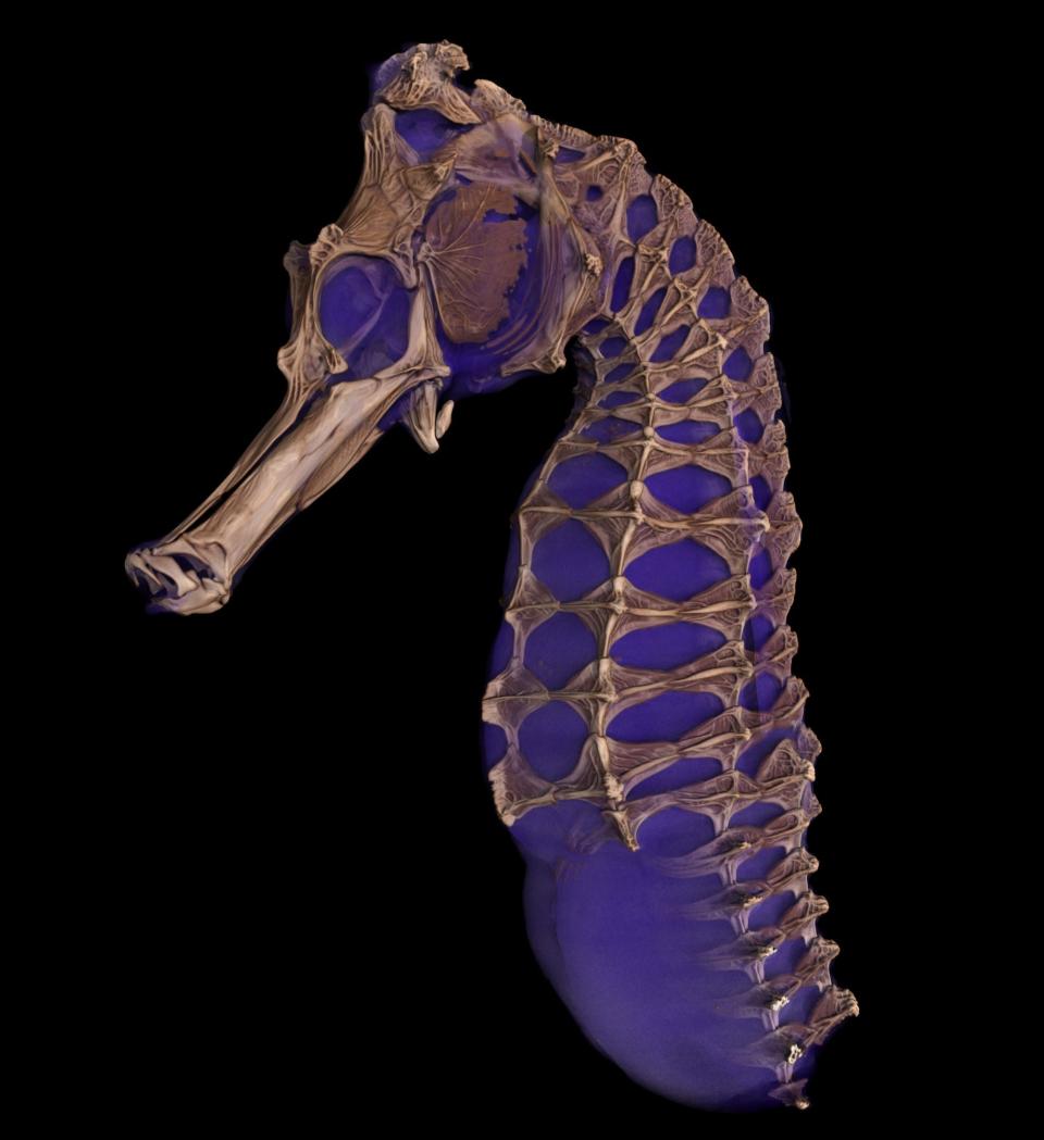
Seahorses look almost the same on the outside as they do in X-rays. This seahorse scan shows the intricate interlocking delicate bones of its skeleton.
Gopher tortoise
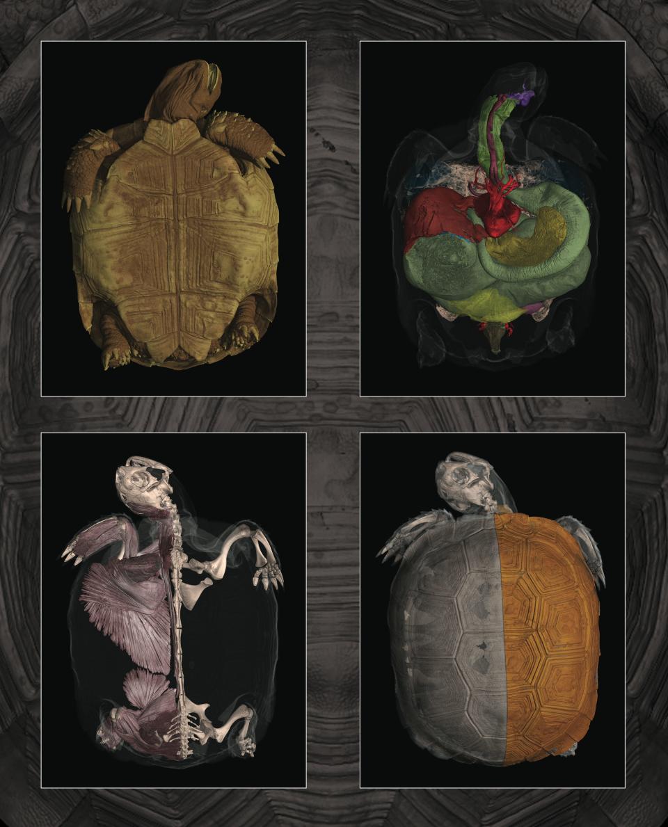
Ever wonder what’s inside a turtle shell? These images show different parts of a gopher tortoise’s anatomy, from its outer shell to its internal organs and skeleton.
3D model of a Komodo dragon skull
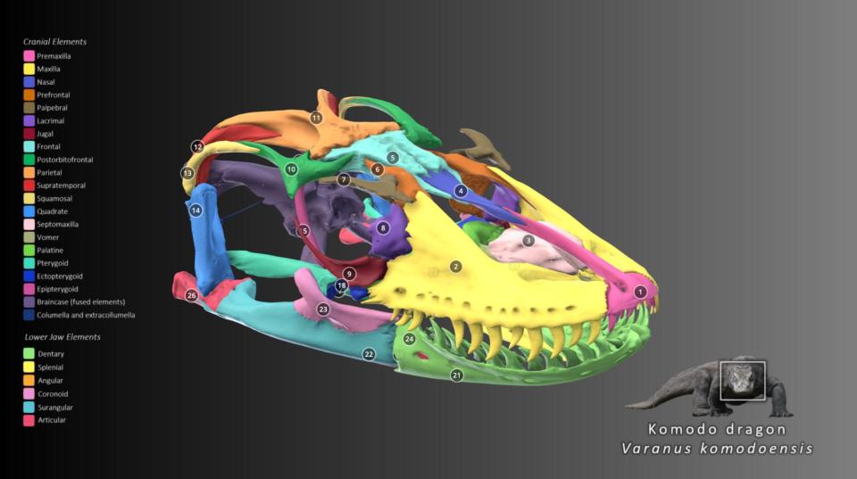
This 3D model of a Komodo dragon’s skull shows its huge jaws, but this lizard has a surprisingly weak grip. Its killing power actually comes from venom stored in a lower jaw gland.
Syrian barefoot buffalo
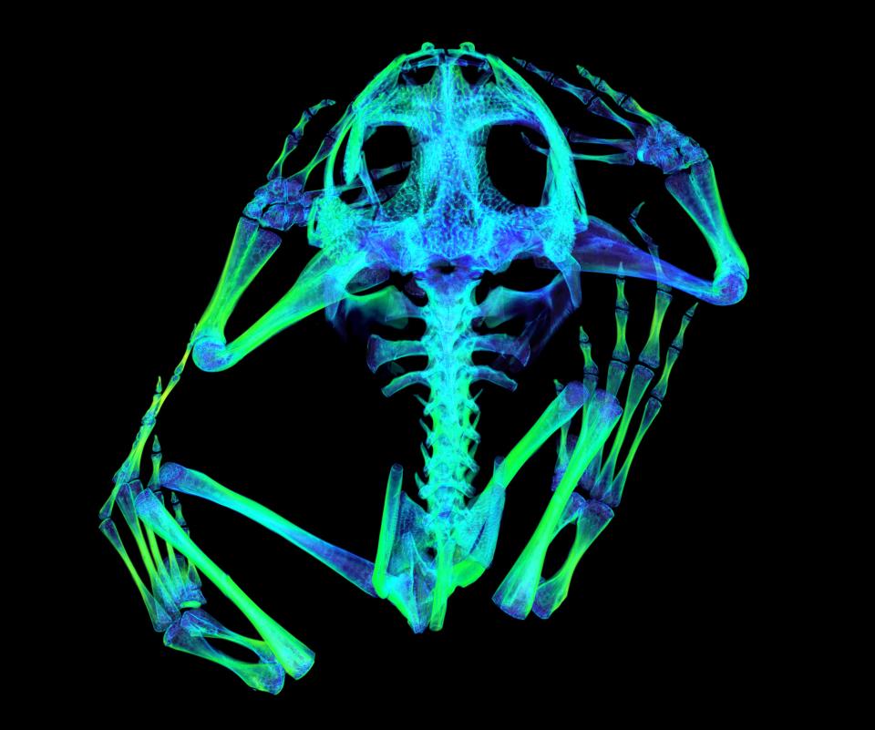
Here’s a color scan of a Syrian spider-footed toad showing its bone density. Green areas are high density and dark blue areas are low density. The low density in the limbs shows spongy bone, which allows for more flexibility.
A mother-lizard and her unborn children
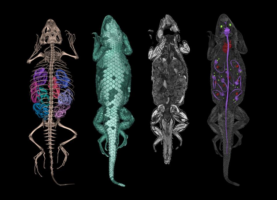
A scan revealed something hidden inside this spiny lizard: babies! On the left, you can see the skeletal structure of this mom lizard and all eight of her offspring. And on the right, you can see the brains, spinal cords, hearts and eyes of the baby lizards.
A pregnant Mexican musk turtle
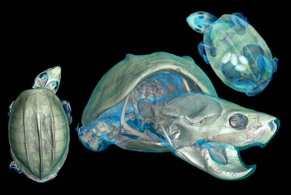
Another surprise was hidden inside this musk turtle specimen from Mexico. Looking closely at the top right image, you will see four eggs inside their coelom.
His turn is green
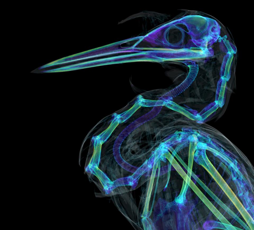
This scan of a green heron showed that unlike other bird species its trachea is much shorter than its neck vertebrae. This indicates that the trachea is very flexible, stretching like an accordion when the hornet extends its long neck to catch prey.
Roloway monkey skull
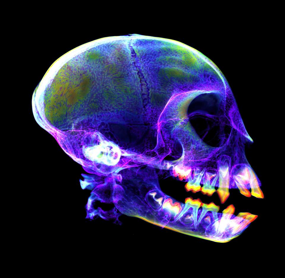
If this image looks familiar, that’s because it’s our monkey cousins. This X-ray of a roloway monkey shows permanent teeth preparing to erupt and replace the specimen’s baby teeth.
Read the original article on Business Insider